
Conference:
CIA-CT on industrial applications of Computed Tomography
11th Conference
19 May 2026
| Technical University of Denmark Meeting Center, building 101, room S09 Anker Engelundsvej 1 2800 Lyngby |
Registration deadline 12 May 2026
About the conference
This year’s conference introduces the most recent advancements in Computed Tomography along with producing insight into diversified applications of CT scanning, ranging from industrial components including medical implants, to building materials, cultural heritage objects, and food products.
Some examples of AI-enhanced CT applications will be presented.
Organizers
The conference is organized by Leonardo De Chiffre, Professor emeritus, DTU Construct, Technical University of Denmark.
Program
|
Registration and light breakfast | |||
|
Welcome and introduction by Venkata Nadimpalli, DTU Construct | |||

|
CT analysis of additively manufactured orthopaedic implant structures (talk is online) Paul Bills, Doctor, University of Huddersfield, Great Britain 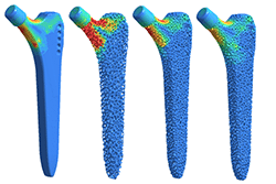 With the growing clinical shift toward uncemented orthopaedic implants, ensuring reliable lattice structures is critical for achieving stable biological fixation. |
|||
|
XCT performance : image quality and dimensional measurement accuracy in standardization Anne-Françoise Obaton, Researcher in metrology for additive manufacturing, LNE, France 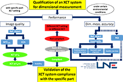 As additive manufacturing (AM) moves toward industrial series production of parts in critical sectors, compliance with geometric and dimensional specifications, within defined tolerances, must be demonstrated. AM enables highly complex geometries with internal structures, making X-ray Computed Tomography (XCT) an essential metrological tool. Although XCT lacks traceability, the growing need for metrology in AM series production implies standardized methodologies, including simplified approaches to measurement uncertainty. |
|||
|
Coffee and networking break | |||

|
Capabilities of AI for automated defect recognition in the X-Ray industry Stephan Tschechne, Dr. Product Manager X-Ray, Carl Zeiss Industrielle Messtechnik GmbH, Germany 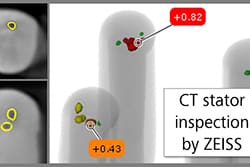 X-Ray technology plays a significant role in Non-Destructive Testing (NDT) of industrial parts across various sectors. |
|||

|
Inspection of drillcores with microCT Sina Maria Baier Stegmaier, Research Engineer, DTU Physics 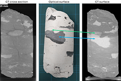 X‑ray microCT is a powerful, non‑destructive technique for examining drill cores, as it reveals their internal structure in 3D without cutting or altering the samples. It enables geoscientists to identify fractures, lithological boundaries, porosity features, and inclusions with high spatial resolution. The resulting datasets also support quantitative analysis—such as density variations or volume fractions—thereby improving geological interpretation. This work explores the application of X‑ray microCT for efficient core logging and highlights key requirements for advancing future applications. Dr. Sina Baier-Stegmaier is a research engineer at DTU Physics and works mainly with industrial application of X-ray CT at the 3D Imaging Center at DTU. She has a background in chemistry and performed her PhD at the Karlsruhe Institute of Technology working with X-ray microscopy and electron microscopy to study heterogeneous catalysts. |
|||
|
Lunch and networking break | |||

|
Industrial applications: From Full Reconstructions to Sparse-Projection Strategies Glenn Gunner Brink Nielsen, Project Manager, Danish Technological Institute  Computed Tomography is increasingly deployed in quality control spanning food processing, agriculture, and wood products. While full CT acquisition remains the gold standard for detailed volumetric analysis, many inline inspection scenarios demand higher throughput than conventional scanning permits. When products come from a known domain with well-characterized material properties, sparse-projection strategies offer a practical path toward fast, scalable inspection. |
|||

|
Sparse-view and dual-energy CT for meat inspection – and other applications Rasmus Juul Pedersen, PhD Student, DTU Compute Dual‑energy X‑ray imaging can address key limitations of sparse‑view CT by providing accurate estimates of known material types along each ray path. Even with few projections, dual‑energy measurements enable separation of materials—such as lean, fat, and bone in meat inspection—and yield reliable per‑ray material distributions. When these distributions are combined across multiple angles and integrated with constraints from complementary vision‑based measurements, it becomes possible to derive parameters such as layer thickness and other internal structure. This can be used for fast, robust inspection of multi‑material objects where full CT acquisition is impractical, including but not limited to industrial meat processing where time is limited. 2025- PhD Student at DTU Compute Section for Visual Computing.2024-2025 Research Assistant at DTU Compute Section for Visual Computing, working on Neural representation for CT reconstruction. 2022-2024 MSc Human-Centered Artificial Intelligence from DTU. |
|||
|
Coffee and networking break | |||

|
Hierarchical Multiscale X-ray Phase-Contrast Tomography: Achieving centimeter-scale context to nanoscale detail in a single optical setup Nis Christian Gellert, Postdoc, DTU Physics  This presentation introduces a hierarchical multiscale X-ray phase-contrast tomography setup developed at DTU and implemented at the DanMAX beamline at MAX IV. The instrument enables imaging of centimeter-scale samples while resolving sub-micron and nanoscale structural details within the same optical configuration. Operating across the full DanMAX energy range (15–35 keV), the system combines a large field-of-view detector with multilayer Laue lenses to bridge the traditional trade-off between resolution and sample size. The approach enables acquisition of multiscale structural information from intact samples, providing both global context and fine internal features. The concept is compatible with higher-energy hard X-rays, making it suitable for dense materials such as minerals, rocks, and structural materials, while maintaining phase-contrast sensitivity for weakly absorbing biological specimens. Nis Christian Gellert is a postdoctoral researcher at DTU Physics working on X-ray phase-contrast tomography and instrumentation development. He holds a PhD from DTU Space and has a background in nanofabrication, X-ray optics, and ray-tracing simulations. His current work focuses on the implementation and development of the XTREME-CT multiscale tomography setup at the DanMAX beamline at MAX IV. |
|||

|
High throughput CT scanning for natural heritage Carsten Gundlach, Senior Research Engineer, DTU Physics We have a vision that high‑throughput X‑ray computed tomography (CT) can enable large‑scale, non‑destructive digitisation of natural heritage collections. |
|||
|
Closing remarks |
Registration fee
| DKK 2,995 | Members of Teknologisk Videndeling and promoting partners listed in the registration form |
| DKK 3,595 | Non-members |
| DKK 1,125 | PhD Students |
| DKK 200 | BSc and MSc students (To register as student you need to have a membership, which is free of charge for students – register here.) |
All prices are excluded of Danish VAT 25%.
The fee includes talks, breakfast, lunch, coffee breaks, refreshments after the seminar and access to speakers’ presentations.
Early bird discount of DKK 300 by registering before 17 April 2026. Early-bird discount does not apply to BSc, MSc and PhD students.
Invoice will be forwarded 14 days before the event. It is possible to pay by credit card.
Cancellation of event
Should we have to cancel the event you will be notified approximately 14 days before the scheduled activity.
Registration
Binding registration
Registration is binding, however substitutions are accepted at any time. Please just contact us at teknologiskvidendeling@construct.dtu.dk.
Questions
Please do not hesitate to contact Teknologisk Videndeling by e-mailing teknologiskvidendeling@construct.dtu.dk
Promoting partners


